
Introduction
Skin and soft tissue infections (SSTIs) are caused by microbial invasions of the skin layers and underlying tissues, because of bacterial, fungal, and viral agents. The current standard of care (CSC) for diagnosing SSTIs involves assessment of clinical history, physical examination, laboratory investigation and surgical examination, delaying optimal and prompt treating. It was recently demonstrated that optical imaging devices (OIDs) could provide alternatives to CSC for the identification of bacterial and fungal infections. Specifically, spectral imaging (SI) techniques including reflectance, fluorescence and Raman spectroscopy imaging are very promising due to the rich set of spectral and spatial information collected by them.
The main relevance of the project results is exploiting spectral imaging technologies to improve accuracy, and speed up the identification of SSTI pathogens in clinical practice. Information obtained by image analysis will help microbiologists to quickly identify a pathogenic microorganism, either in a Petri dish, in a film on a medical device or on a microscopic slide. In the clinical setting the results of the project will show if spectral imaging is a feasible technique for real time in-vivo diagnostics of SSTI.
Summary
The main objective of the proposed research is to facilitate translation of optical imaging devices into clinical practise and Full Laboratory Automation (FLA) for a faster and more accurate identification and differentiation of skin and soft tissue infections (SSTI) microbes. Specific objectives of the research, including a brief work description, are: (1) Spectral imaging of SSTI microbe cultures. Microbial cultures of common SSTIs will be imaged using VIS-NIR reflectance, fluorescence and Raman spectral imaging (SI). Different substrates will be used for microbial cultivation, simulating FLA environment and real skin infections. (2) Spectral imaging of SSTI in-vivo. Patients with SSTI will be imaged using SI. (3) Development of standardised image processing pipelines. Images compiled in the database created under objective (4) will be used to create automated image processing pipelines (IPPL) to achieve optimal sensitivity and specificity for identification and differentiation of SSTI microbes for FLA and clinical inspection. (4) Creation of a publicly available database of SI images. An online database environment will be prepared for the storage, access, processing and exchange of spectral images of SSTI microbial cultures (1) and in-vivo images (2). The project will involve researchers having different backgrounds: medical hyperspectral imaging, microbiology, medical image processing and clinical dermatology. Participating institutions will provide the instrumental, microbial culturing, image processing capabilities, storage infrastructure and patient access needed for the proposed research. The results of this research, including the standardisation of spectral image processing pipelines, will enable a smoother transition of the technology into clinical practise. The database of spectral images developed will enable further development of image processing algorithms by the wider scientific community.
Methodology
The current standard of care (CSC) for diagnosing SSTI involves assessment of clinical history, physical examination, laboratory investigation and surgical examination. Laboratory investigation provides microbiology reports identifying the pathogens, but these details are not provided immediately, but rather in a couple of days or weeks. Therefore, faster and reliable means of SSTI diagnosis are needed. The main objective of the proposed research is to facilitate translation of optical imaging devices (OID) – which showed promising results for specific SSTIs – into clinical practice and full laboratory automation (FLA) for a faster and more accurate identification and differentiation of SSTI microbes. The main problems of OID translation to clinical workflow are: (1) lack of a standardised end-to-end automatic image processing pipeline, (2) lack of standardised image acquisition parameters, (3) neglecting colony/infected tissue morphological information, (4) lack of substrate variations, and (5) lack of publicly available spectral images of SSTI colonies and infected tissues.
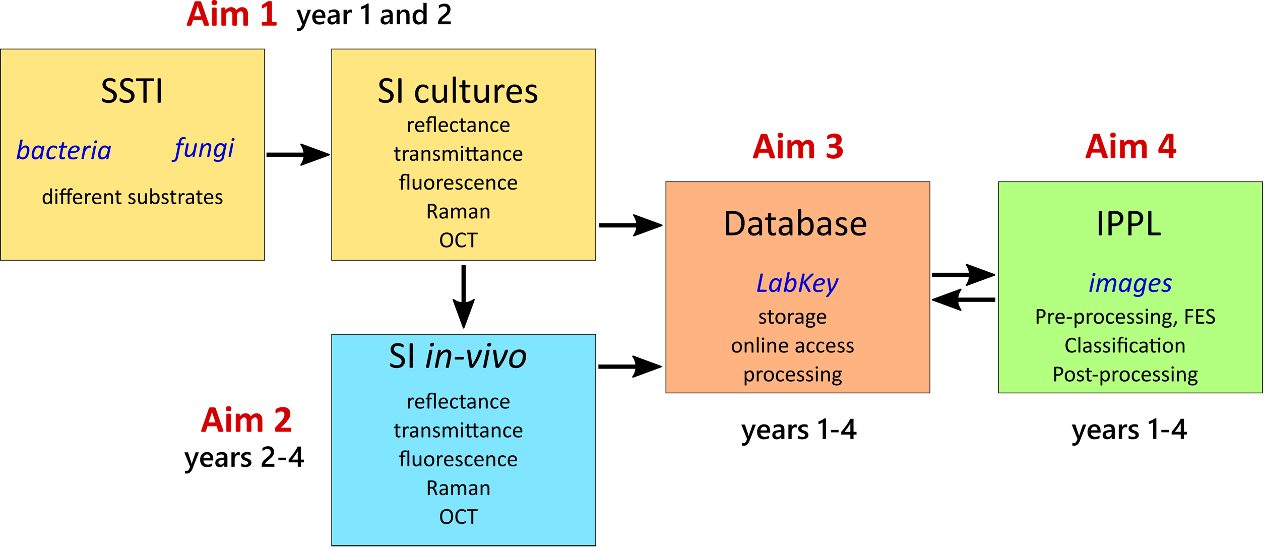
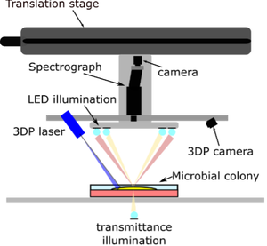
The research methodology will include: (1) spectral imaging of SSTI microbe cultures. Microbe cultures of common SSTIs will be imaged using VIS-NIR, fluorescence and Raman SI, and OCT imaging – all combined in a laboratory imaging system. (2) Spectral imaging of SSTI in-vivo. Patients with SSTI will be imaged using a custom developed SI and OCT systems. (3) Development of end-to-end image processing pipelines based on the obtained images. The images assembled in the database created under the objective (4) will be used in creating image processing pipelines (IPPL) for obtaining optimal sensitivity and specificity of the SSTI microbe identification and differentiation for FLA and clinical inspection. (4) Creation of a publicly available database of SI images. An online database environment for storing, accessing, manipulating and sharing spectral images of SSTI microbe cultures (1) and in-vivo images (2) will be prepared.
Results
The most significant project results are: (1) a collection of reflectance, transmittance, fluorescence, Raman, and OCT images of the microbe cultures; (2) a collection of reflectance, transmittance, fluorescence, Raman and OCT images of the SSTIs in-vivo; (3) validated IPPLs for obtaining optimal sensitivity and specificity of the SSTI microbe identification and differentiation; (4) a functioning publicly accessible database of standardised SI images, algorithms and infrastructure for sharing datasets and algorithms by researchers and industry.
Research Group
prof. Ivan Štajduhar 1 – principal investigator (Croatian team)
prof. Matija Milanič 3 – principal investigator (Slovenian team)
prof. Marija Kaštelan, M.D. 2
prof. Larissa Prpić Massari, M.D. 2
prof. Martina Modic 4
prof. Urban Simončič 3
Marijana Vičić, M.D., M.Sc. 2
Nika Hlača, M.D. 2
Tina Žagar, M.D. 2
Jošt Stergar, Ph.D. 4
Franko Hržić, Ph.D. 1
Dominik Vičević, mag. ing. comp. 1
Mateo Mikulić, mag. ing. comp. 1
Tadej Tomanič 3
Rok Hren 3
& associates
1 University of Rijeka, Faculty of Engineering
2 University of Rijeka, Faculty of Medicine / Clinical Hospital Centre Rijeka
3 University of Ljubljana, Faculty of Mathematics and Physics
4 Jožef Stefan Institute, Ljubljana
Resources
Our image repository can be found on https://optiderm.riteh.hr/ (currently under development)
Funding
Funded by Croatian Science Foundation grant IP-2022-10-2433 and Slovenian Research Agency grant N3-0348.